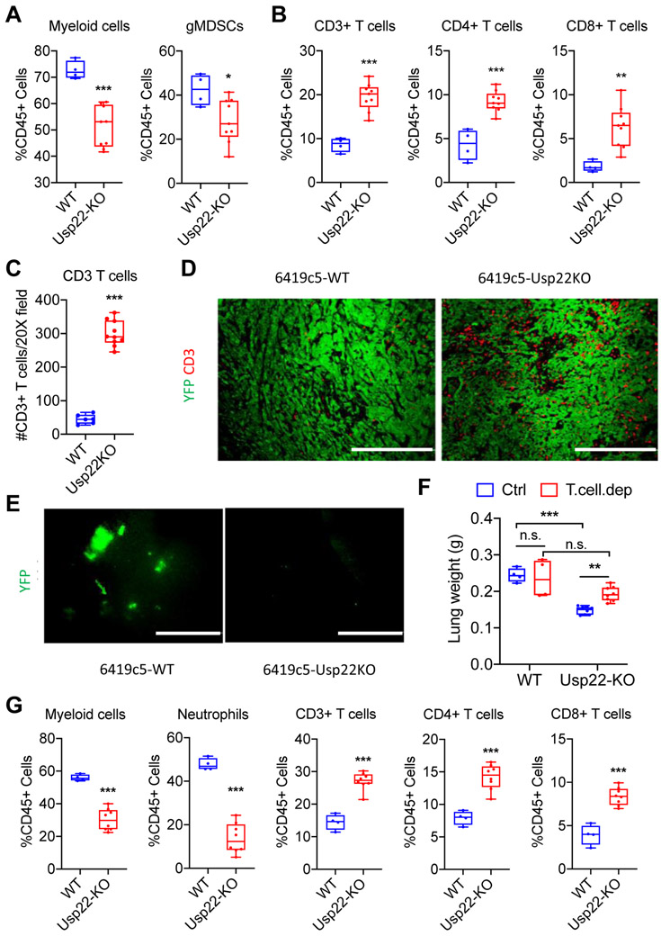
Figure 2. Loss of tumor cell–intrinsic USP22 promotes antitumor immunity in orthotopic PDA tumors and suppresses lung metastasis.
(A-B) Flow cytometric analysis of tumor-infiltrating immune cells in orthotopically implanted Usp22-WT and Usp22-KO tumors (2 independent knockout clones, n=4-9 tumors analyzed per group in 2 experiments). (C) Quantification and (D) representative immunofluorescent staining of CD3+ T cells in Usp22-WT and Usp22-KO orthotopic tumors (2 independent knockout clones, n=6-10 tumors analyzed per group) stained for CD3 (red) and YFP (green). (E) Representative images of lungs from animals in (D) with YFP+ tumor cells shown in green. (F) Quantification of lung weight 14 days after tail vein injection of Usp22-WT or Usp22-KO tumor cells (2 independent knockout clones) with or without T cell depletion (n=5 mice/group). (G) Flow cytometric analysis of lungs following tail veil injection of Usp22-WT or Usp22-KO tumor cells (2 independent KO clones, n=5-10 mice/group in 1 experiment). In (A-C & F-G), data are presented as boxplots with horizontal lines and error bars indicating mean and range, respectively. In (A-C & G), statistical differences between groups were calculated using Student’s unpaired t-test. In (F), statistical differences between groups were calculated using 2-way ANOVA analysis with multiple comparison. *p<0.05, **p<0.01, *** p< 0.001. Scale bars, 200 μm in (D), 1 mm in (E).

