
Figure 1.
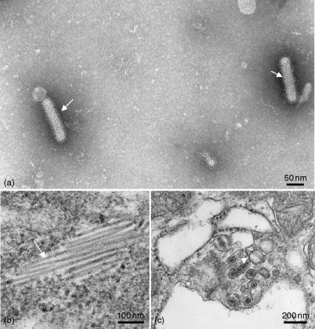
Transmission electron micrographs of YHV. (a) Image of virions stained with heavy metal salts showing the external appearance of the enveloped particles (arrows). (b) Image of an ultrathin section of helical nucleocapsids (arrow) within the gill of an infected shrimp. (c) Image of an ultrathin section of virions (arrow) within the gill of an infected shrimp. Kindly provided by Dr. Alex Hyatt, CSIRO, Australian Animal Health Laboratory, Geelong, Australia.