
Figure 2.
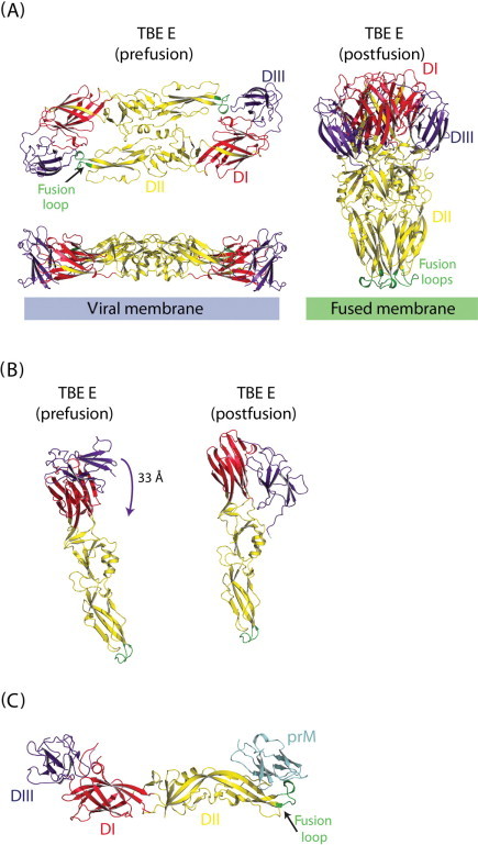
Class II fusion proteins. (A) TBEV glycoprotein E structures. Ribbon diagram of the pre-fusion E dimer (Rey et al., 1995) (left part) and post-fusion E trimer (Bressanelli et al., 2004) (right part). Red yellow and blue part of the protein correspond to respectively the central domain (DI), the dimerization domain (DII) and the C-terminal domain (DIII); fusion loop is depicted in green and located at the tip of DII. (B) TBEV E protomer conforlmational change. Ribbon diagram of the pre- and post-fusion E protomer of TBEV. The arrow indicates the movement of domain III (connected to the transmembrane domain of the protein) toward the fusion loop. (C) Dengue virus precursor membrane-envelope protein complex (prM–E) structure. Ribbon diagram of the prM-E heterodimer (Li et al., 2008). The pr peptide, in light blue, protects the fusion loop in green. Domains of E are indicated and colored according to A. (See Color Insert.)