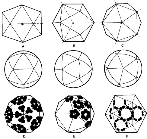
FIG. 1-2.

Features of icosahedral structure. A regular icosahedron viewed along twofold (A), threefold (B), and fivefold (C) axes of symmetry. In negatively stained electron micrographs, virions may appear hexagonal in outline (upper row) or apparently spherical (middle row). Various clusterings of capsid polypeptides give characteristic appearances of the capsomers in electron micrographs (lower row). For example, they may be arranged as 60 trimers (D), capsomers being then difficult to define, as in poliovirus; or they may be grouped as 12 pentamers and 20 hexamers (E), which form bulky capsomers as in parvoviruses; or as dimers on the faces and edges of the triangular facets (F), producing an appearance of a bulky capsomer on each face, as in caliciviruses.
