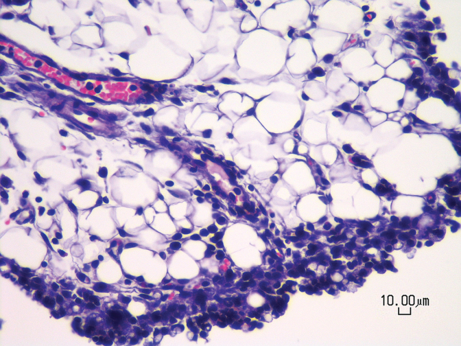
FIG. 5.
Photomicrograph of a vehicle-treated mouse showing mild to moderate chronic peritonitis characterized by the presence of lymphocytes, plasma cells, and macrophages within peritoneal fat. All animals receiving daily IP injections for 5 days (n=4–9/treatment) exhibited the same finding regardless of treatment with drug or vehicle. Hematoxylin and eosin stain, 400× magnification, scale bar=10 μm. IP, intraperitoneal.

