
Figure 4.
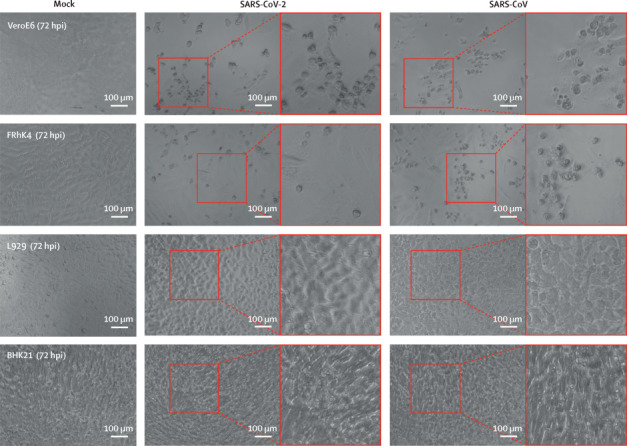
Detection of SARS-CoV-2-induced cytopathic effects in representative cell types
SARS-CoV-2-induced cytopathic effects were assessed in VeroE6 and FRhK4 cells. L929 and BHK21 cells were included as negative controls. Cells were infected with SARS-CoV-2 or SARS-CoV at 0·1 MOI. At 72 hpi, typical cytopathic effects were seen, including cell rounding, detachment, degeneration, and syncytium formation. Boxed area is shown adjacent to each image. Cells were imaged with a Nikon Ts2R-FL inverted microscope. Bars represent 100 μm. SARS-CoV-2=severe acute respiratory syndrome coronavirus 2. SARS-CoV=severe acute respiratory syndrome coronavirus. MOI=multiplicity of infection. hpi=hours postinoculation.