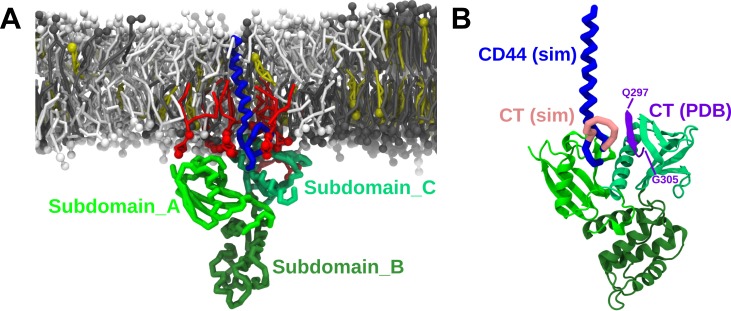
Fig 7. PIP2-dependent formation of CD44-FERM complex.
(A) Presentation of CD44-FERM conformation and association on the membrane surface in the presence of PIP2. The backbone structure of CD44 is shown in blue, the FERM domain is shown in different shades of yellow/green, dependent on the subdomain. The snapshot in a bottom-view is exhibited in the left-upper corner. B) Comparison of the binding mode from our simulation with the crystal structure of the FERM-CT complex. The FERM domain of both structures are aligned. The CT domain (Q297-G305) in the crystal structure is colored in purple, and in pink in our simulation.

