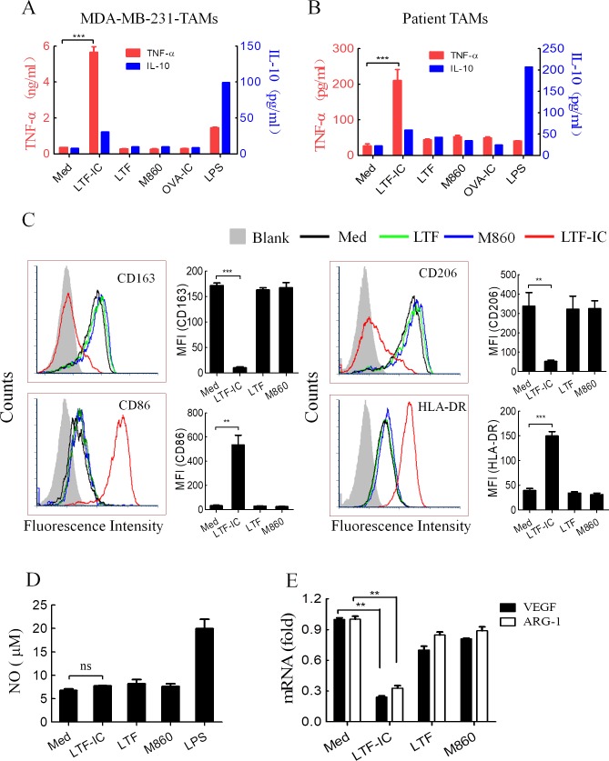
Figure 1.
LTF-IC resets human TAMs to M1 phenotype. MDA-MB-231-TAMs derived from human peripheral blood monocytes (A) or macrophages purified from ascites of patients with breast cancer using CD14 microbeads (B) were treated in triplicate wells with LTF, M860, LTF-IC, OVA-IC (30 µg/mL) or LPS (100 ng/mL) for 24 hours and then the culture supernatants were collected for quantification of TNF-α and IL-10 using ELISA kits. Unstimulated cells were also included as negative control (Med). (C) Following 24 hours treatment with, or without (Med), LTF, M860 or LTF-IC (30 µg/mL), MDA-MB-231-TAMs were analyzed for surface expression of CD163, CD206, CD86 and HLA-DR using FACS. Representative histograms are presented, mean fluorescence intensity (MFI±SD) of cells from triplicate wells are shown in bar graphs on the right. (D) No concentration (mean±SD) in the culture supernatant of MDA-MB-231-TAMs after 24 hours stimulation with, or without (Med), LTF-IC, LTF, M860, or LPS was quantitated using the NO colorimetric assay kit. (E) VEGF and ARG-1 mRNA levels in MDA-MB-231-TAMs after 24 hours stimulation with, or without (Med), LTF-IC, LTF or M860 in triplicate wells were detected using Q-PCR. The results (mean±SD), after normalization to GAPDH mRNA expression, are expressed as fold increase compared with the Med group. The results are representative of at least three experiments using cells from different donors. *P<0.05, **P<0.01, ***P<0.001. ARG-1, arginase-1; IC, immunocomplex; IL-10, interleukin-10; LPS, lipopolysaccharide; LTF, lactoferrin; OVA, ovalbumin; Q-PCR, quantitative PCR; TAMs, tumor-associated macrophages; TNF-α, tumor necrosis factor α; VEGF, vascular endothelial growth factor.

