. 2019 Apr 2;16(2):338–340. doi: 10.3988/jcn.2020.16.2.338

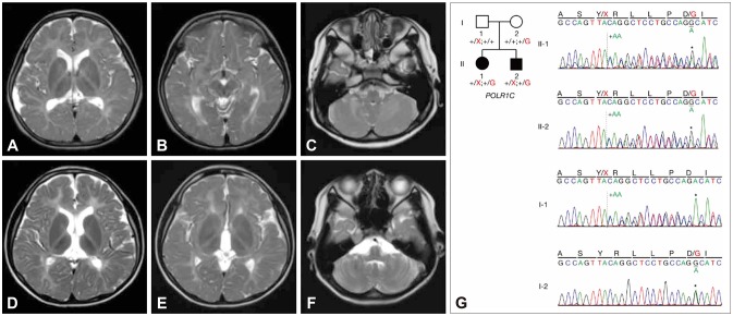
Fig. 1. Neuroimaging (A–F) and Sanger sequencing (G) findings. Besides diffuse hypomyelination, relative T2-weighted MRI hypointensities in the ventrolateral thalamus and optic radiation are evident in the proband (A and B) and her affected brother (D and E). Hypomyelination of the cerebellar white matter is visible, without cerebellar atrophy (C and F). (G) Compound heterozygous variants in POLR1C are shown in the pedigree: a frameshift mutation (c.698_699insAA: p.Tyr233fs) and a missense mutation (c.713A>G: p.Asp238Gly). Solid symbols indicate clinically affected and genetically confirmed patients: the proband (II-1) and the affected brother (II-2). One variant was inherited from their mother, the other from their father.

Fig. 1