Figure 1.
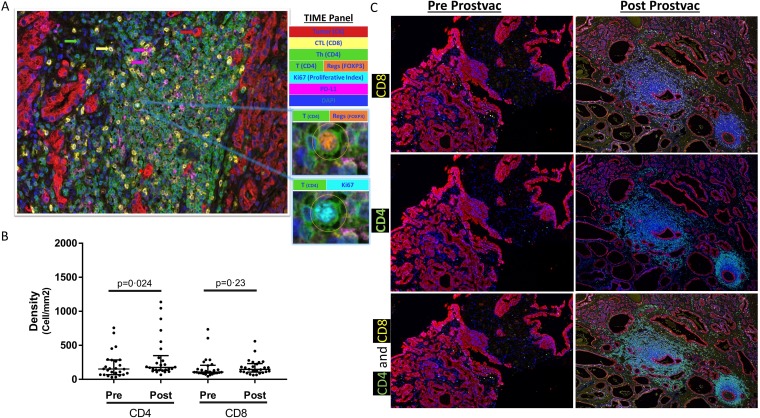
Cell densities in prevaccination biopsies (Pre) and in radical prostatectomy sections postvaccination (Post) with PROSTVAC. (A) Representative image of multiplex immunofluorescence panel. Insets show a cell expressing four markers (CD4, FOXP3, Ki67 and DAPI). (B) Immune-cell infiltrates were quantified in both prevaccination and postvaccination sections using inForm software. NCA of both CD4 and CD8 immune-cell density ratios was assessed by the Wilcoxon signed rank test. Median±IQR shown with horizontal lines. (C) Exceptional case with CD4 and CD8 immune-cell infiltrates prevaccination and postvaccination. CTL, cytotoxic T lymphocyte; NCA, non-compartmentalized analysis; PD-L1, programmed death-ligand 1; Th, T helper; TIME: tumor immune microenvironment.

