Figure 5.
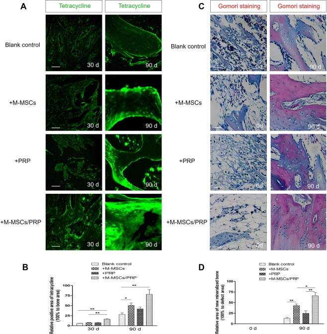
M-MSCs/PRP gel significantly improved the formation of new bone tissues. (A,B) Compared with blank control group, tetracycline fluorescence signal was not significantly enhanced in PRP group until 90 d; both M-MSCs and M-MSCs/PRP group displayed apparent tetracycline fluorescence signal. Compared with M-MSCs single group, M-MSCs/PRP group displayed stronger tetracycline fluorescence signal. Scale bar = 100 μm. (C,D), The new mineralized bone was labeled “red” by Gomori staining. Compared with blank control group, M-MSCs group or PRP group, the new mineralized bone area was significantly increased in M-MSCs/PRP group at 90 d. Scale bar = 100 μm. 3 randomized fields of view for each group, *P < 0.05, **P < 0.01.

