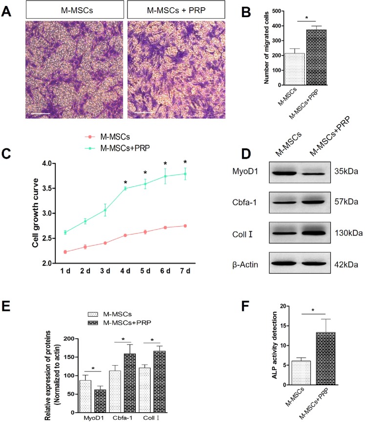
Figure 7.
PRP treatment promotes migration, proliferation and induced osteogenic differentiation of M-MSCs in vitro. (A,B) Transwell assay displayed that after PRP treatment, the number of migrated M-MSCs was significantly increased. 10 randomized fields of view for each group. scale bar = 20 μm. (C) CCK8 assay displayed that after PRP treatment, cellular growth was significantly enhanced. (D,E) Western blot analysis displayed compared with M-MSCs treated with FBS, PRP treatment increased the expression of Cbfa-1 and Coll I while decreased the expression of MyoD1. (F) ALP activity detection revealed PRP treatment significantly elevated ALP activity in differentiated M-MSCs. *P < 0.05, **P < 0.01.

