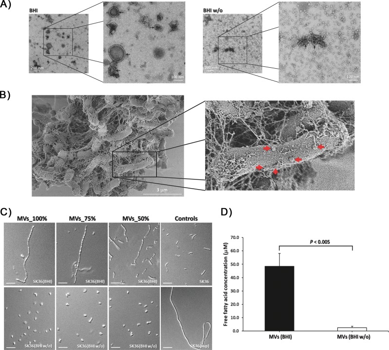
Fig. 6. Transmission electron micrograph of MVs prepared from Cd.
a 1:10-diluted samples in low and high magnification isolated from Cd grown in BHI (left) and BHI w/o (right), showing the differences of double-walled vesicle-like structures. Arrows indicate spherical MVs. b SEM image of Cd cells cultured in biofilm conditions showing spherical blebs (arrows) surrounding the bacteria. The pictures are representative of three independent experiments. Scale bars are indicated on each panel. c SK36 chain length morphology after being treated with MVs prepared from Cd grown in BHI and BHI w/o compared with Cd supernatants (sup) and the BHI controls. d Average chain length (μm). Data are presented as the means of three biological replicates. Error bars denote standard deviations. Scale bars indicate 10 μm.

