Figure 3.
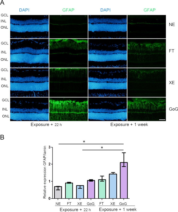
GFAP stress response after a total dose of 0.5 J/cm2. Male Wistar rats aged 7 weeks were exposed to GaN-on-GaN (GoG) LED or conventional white LED(XE) (Xanlite XXX Evolution 5 W) or fluorescent tubes (FT) at 1900 lx, receiving the estimated retinal dose of 0.5 J/cm2 (2 h for GoG, 2h20 min for XE and FT). 22 hours or 1 week after the end of the exposure period (A) the eyes were included in optimal cutting temperature medium (Tissue Tek), cryosectioned and immunostained with anti-Glial Fibrillary Acid Protein (GFAP) (green). NE: Non exposed. Nuclei were stained in blue with DAPI. ONL indicates the outer nuclear layer, INL the inner nuclear layer and GCL the ganglion cell layer. Scale bar represents 100 µm. The presented photographs were taken in the superior part of the retina, at 200 µm from the optic nerve. (B) the eyes were enucleated, the retina dissected, extracted with M-PER buffer and loaded on the top of a 10% SDS-PAGE, transferred onto a nitrocellulose membrane and probed with anti-GFAP. Lamin B was used as a charge control. Histograms represent the median with the interquartile range. Significance was evaluated using the Kruskal-Wallis test followed by the Dunn’s multiple comparison post-test. H(7) = 19.09. *p ≤ 0.1, n = 3.

