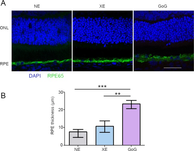
Figure 8.
RPE damages induced by a total retinal dose of 2.2 J/cm2. Male Wistar rats aged 7 weeks were exposed to GaN-on-GaN (GoG) LED or conventional white LED (XE) (Xanlite XXX Evolution 5 W) for 9 and 10 h repectively at 1900 lx, receiving the estimated retinal dose of 2.2 J/cm2 (BLH weighted 0.253 J/cm2 for regular LED and to 0.26 J/cm2 for GaN-on-GaN LED). (A) 15 hours after the end of the exposure period the eyes were included in optimal cutting temperature medium (Tissue Tek), cryosectioned and immunostained with anti-retinal pigment epithelium protein 65 (RPE 65). Nuclei are stained with DAPI in blue. ONL indicates the outer nuclear layer, RPE the retinal pigment epithelium. Scale bar represents 100 µm.NE: Non Exposed. (B) Measurement of the RPE thickness after exposure. Histograms represent the median with the interquartile range. Significance was evaluated using the Kruskal-Wallis test followed by the Dunn’s multiple comparison post-test. H(3) = 26.28. ***p ≤ 0.001, n = 12.

