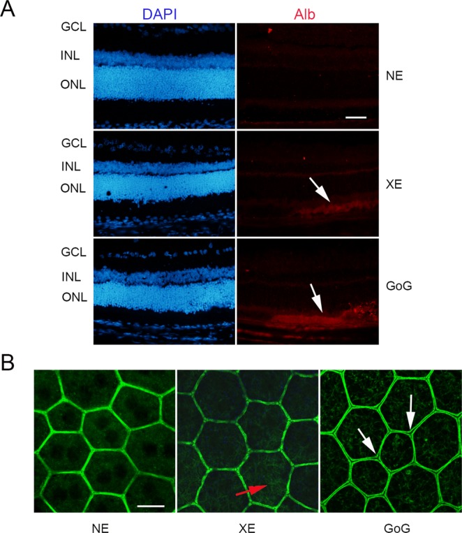
Figure 9.

The RPE after a total retinal dose of 2.2 J/cm2. Male Wistar rats aged 7 weeks were exposed to GaN-on-GaN (GoG) LED or conventional white LED (XE) (Xanlite XXX Evolution 5 W) for 9 and 10 h respectively at 1900 lx, receiving the estimated retinal dose of 2.2 J/cm2 (BLH weighted 0.253 J/cm2 for regular LED and to 0.26 J/cm2 for GaN-on-GaN LED). (A) 15 hours after the end of the exposure period the eyes were included in optimal cutting temperature medium (Tissue Tek), cryosectioned and immunostained with anti-Rat seric albumin (alb). White arrows indicate albumin leakage. ONL indicates the outer nuclear layer, INL the inner nuclear layer and GCL the ganglion cell layer. NE: Non exposed. Scale bar represents 100 µm. (B) alternatively the eyes were dissected and the posterior pole containing the RPE and the choroid was flat mounted, stained with labeled phalloidin (green) and analyzed under a confocal microscopy. Red arrow indicates stress fibers, white arrows show interruptions of the OBRB. Scale bar represents 10 µm.
