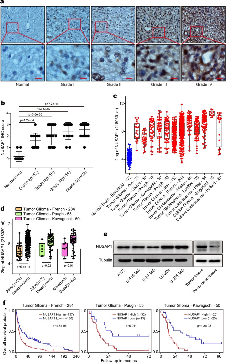
Fig. 1.
NUSAP1 is highly expressed in GBM patients and in GBM cells. a Immunohistochemical analyses of NUSAP1 in normal brains and glioma tissues. b Statistical analyses of NUSAP1 in 8 normal brains and 64 glioma tissues. c The expression of NUSAP1 in normal brain as well as in glioma patients and in cell lines from 13 different data sets. d Analyses of NUSAP1 in living patients and in dead patients from three different databases. e The level of NUSAP1 in five GBM cell lines as well as in a pair of peritumoral and tumor tissues. f Kaplan–Meier analysis of progression-free survival using data from three different glioma databases

