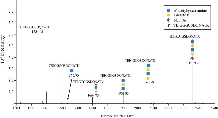
FIGURE 3.
LC-MS/MS analysis of sialylated glycoproteins. Purified FN3 carrying the sialylated glycan was analyzed by LC-MS/MS after enzymatic digestion. The fragmentation spectrum of the Neu5Ac-α-2,6-Gal-β-1,4-GlcNAcβ-1,3-Gal-β-1,3-GlcNAc-TEIGGGGSDQNATK glycopeptide is shown. The peaks at m/z 1334.62 and 2355.98 correspond to the TEIGGGGSDQNATK peptide and the sialylated TEIGGGGSDQNATK glycopeptide containing a NeuAc(1)Hex(2)HexNAc(2) glycan, respectively. Peaks at m/z, and 2064.88, 1902.83, 1699.75, and 1537.70 correspond to loss of, NeuAc(1), NeuAc(1)Hex, NeuAc(1)Hex(1)HexNAc(1), and NeuAc(1)Hex(2)HexNAc(1), respectively, from the sialylated glycopeptide. Blue squares, yellow circles and purple diamonds indicate GlcNAc, Gal, and Neu5Ac residues, respectively.

