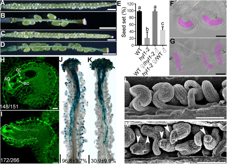
FIGURE 4.
hyl1-2 is defective in ovule development, similar to hen1-8. (A–D) Representative silique of wild type (A), hyl1-2 (B), wild type pollinated with hyl1-2 pollen (C), or hyl1-2 pollinated with wild-type pollen (D). (E) Quantitative analysis of seed set. Results are means ± SD (n = 15). Different numbers indicate significantly different groups (One-Way ANOVA, Tukey’s multiple comparisons test, P < 0.05). (F,G) Whole-mount ovule clearing of wild type (F) or hyl1-2 (G). Embryo sacs are highlighted with lilac. (H,I) CLSM of wild-type (H) or hyl1-2 (I) ovules at stage 3-VI. Ovules were stained with PI and mid-optical sections are shown. Numbers on the bottom of each image indicate displayed ovules/total ovules examined. cc, central cell; ec, egg cell; sg, synergid cell. (J,K) Histochemical GUS staining of wild-type (J) or hyl1-2 (K) pistils at 12 HAP with ProLAT52:GUS pollen. Two to three overlapping high-magnification images were taken for one pistil and overlaid with Photoshop (Adobe) to show the whole pistil. Numbers at the bottom are quantification of targeted ovules out of total ovules. Results are means ± SD (n = 15). (L,M) SEMs of mature ovules from wild type (L) or from hyl1-2 (M). Arrowheads point at ovules with protruding embryo sac. Bars = 1 mm for (A–D); 50 μm for (F,G); 10 μm for (H–I); 200 μm for (J,K); 100 μm for (L,M).

