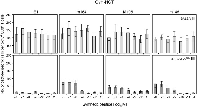
Figure 6.
Quantitation of GvH-reactive and viral epitope-specific liver-infiltrating CD8+ T cells after GvH-HCT. Liver-infiltrating CD8+ T cells were isolated from liver tissue (yield from 10 livers) of infected GvH-HCT recipients on day 20. Their functional activity was tested in an ELISpot assay based on IFNγ secretion in response to stimulation with BALB/c (upper panels) or BALB/c-H-2dm2 (lower panels) MEF as target cells that were exogenously loaded with graded loading-concentrations of the indicated synthetic viral peptides to reveal cumulative avidity distributions. Effector cells responding at a certain peptide concentration include cells that respond to concentrations < the indicated concentration. Ø, no viral peptide loaded. Error bars represent 95% confidence intervals determined by intercept-free linear regression.

