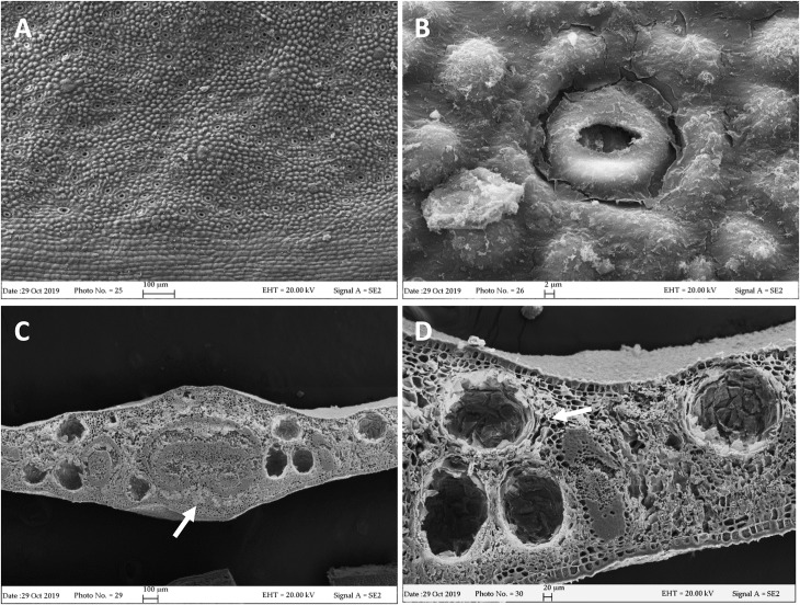
FIGURE 2.
SEM micrographs of E. gunnii leaf. (A,B) Epidermal surface showing papillose cells and many anomocytic stomata. (C) Leaf trasversal section with secretory cavities scattered throughout the mesophyll, around the midrib. (C,D) Prismatic crystals and druses are visible near and around the secretory cavities and the rib (arrows).

