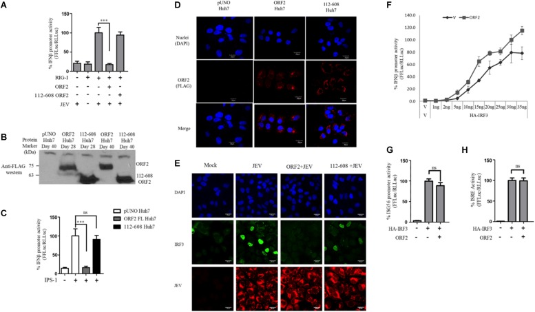
FIGURE 5.
IRF3 nuclear translocation is inhibited by the HEV ORF2 protein: (A) RIG-I assay was performed by co-transfecting RIG-I (0.5 ng) and FL ORF2 or 112-608 ORF2 plasmids with IFN-β firefly and CMV Renilla luciferase reporter plasmids in Huh7 cells. JEV infection was given at the 0.5 MOI for 4 h. Luciferase activity was measured 16 h post-infection. (B) Stable expression of FL ORF2 and 112-608 ORF2 proteins was monitored by western blot analysis. Cell lysates at day 28 and day 40 for each of ORF2 Huh7 and 112-608 Huh7 were probed with anti-FLAG antibody. (C) For IPS-1 assay, IPS-1 (5 ng) and IFN-β firefly and CMV Renilla luciferase reporter plasmids were transfected in pUNO Huh7, ORF2 Huh7, and 112-608 Huh7 stable cell lines. IFN-β promoter activity was measured 24 h post-transfection. (D) pUNO Huh7, ORF2 Huh7 and 112-608 Huh7 cells were treated with the goat anti-FLAG primary antibody and anti-Goat Alexa-568 (red) secondary antibody to visualize the ORF2 proteins. Nuclei were stained with DAPI. The scale represents 20 μm. (E) pUNO Huh7, ORF2 Huh7, and 112-608 Huh7 cells were infected with the JEV at 0.5 MOI for 24 h. Nuclei were stained with DAPI, IRF3 was stained with rabbit anti-IRF3 primary and anti-rabbit Alexa-488 (green) antibody and JEV was stained with mouse anti-JEV glycoprotein E antibody and anti-mouse Alexa-596 (red) antibody. (F) IRF3 assay was performed by co-transfecting increasing quantities of HA-IRF3 and 25 ng of FL ORF2 plasmids with IFN-β firefly and TK Renilla luciferase reporter plasmids. Luciferase activity was measured 24 h post-transfection. (G) IRF3 assay was performed by co-transfecting HA-IRF3 and 25 ng of FL ORF2 plasmids with ISG56 firefly and TK Renilla luciferase reporter plasmids. Luciferase activity was measured 24 h post-transfection. (H) IRF3 assay was performed by co-transfecting HA-IRF3 and 25 ng of FL ORF2 plasmids with ISRE firefly and TK Renilla luciferase reporter plasmids. Luciferase activity was measured 24 h post-transfection. Values are mean ± SD, n = 3 for all experiments. (∗∗∗ denotes p-values ≤ 0.001, ns denotes p-values ≥ 0.05).

