
Figure 5.
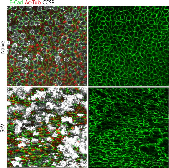
SeV infection alters the normal airway epithelial mosaic. Whole mount immunofluorescence images of proximal portions of the axial bronchi of mice infected or not with SeV 5 weeks earlier (N = 5 mice). Images on the left show staining for E‐cadherin (E‐Cad, green) to outline cell borders, acetylated tubulin (Ac‐Tub, red) to identify ciliated cells and club cell secretory protein (CCSP, white) to identify secretory cells. Images on the right only show staining for E‐cadherin to better illustrate changes in cell shape and pattern. Scale bar = 20 μm