
Figure 1.
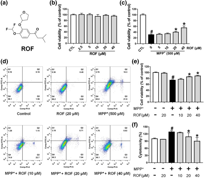
Roflupram increases cell viability of MPP+‐induced cells. (a) The chemical structure of roflupram (ROF). (b) SH‐SY5Y cells were treated with various concentrations of roflupram for 48 hr to evaluate the cytotoxic effects on cells. (c) The cells were pretreated with different concentrations of roflupram for 1 hr, and then treated with 500 μM of MPP+ for 48 hr to measure cell viability with MTT assay. (d and e) The cells were pretreated with various concentrations of roflupram 1 hr earlier and were then exposed to 500 μM of MPP+ for 24 hr; cell viability was measured using flow cytometry. Representative graphs of flow cytometric outputs for each group are shown in (d), and statistical analysis of flow cytometry is presented in (e). (f) The cells were treated with various concentrations of roflupram for 1hr and were then stimulated with 500 μM of MPP+ for 48 hr to measure LDH activity in the medium. Data are presented as mean ± SD (n = 5) and represent five independent experiments. # P < .05, significantly different from control group. *P < .05, significantly different from MPP+‐treated group