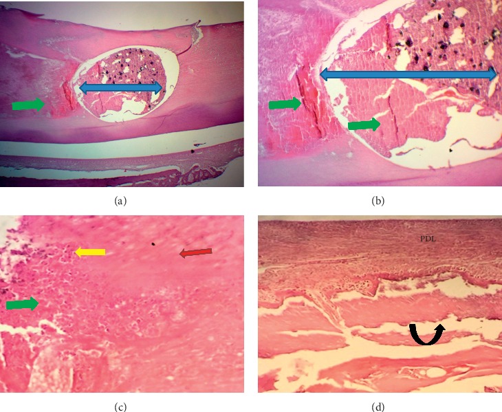
Figure 3.

Microphotographs of an H&E-stained longitudinal section for the pulp and periodontium response in the central incisor after 1 week of MM-MTA treatment (blue left-right arrows). (a) (40X) and (b) (100X): evidence of inflammation, necrosis (green arrows) and calcification within the pulp. (c) Irregular dentine (red arrow) deposition (100X). (d) The periodontal ligament had unorganized fibrous tissue and congested capillaries with inflammatory cells infiltration (yellow arrow, 40X). Small woven bone trabeculae (black curved-up arrow) presented at the interface with PDL.
