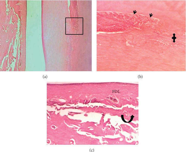
Figure 5.

Microphotographs of an H&E-stained longitudinal section through central incisor after 4 wks of Biodentine (BD) treatment. (a-b) A highly cellular pulp tissue obliterated by irregular deposition of reparative dentine (quad arrow) next to multiple layers of small-sized odontoblasts “Black arrow heads” (40X and 100X). (c) The periodontal ligament attached to a thin cementum layer had organized and dense fibrous bundles running parallel to the root surface (principle fibers) besides a few congested and dilated capillaries toward the bone side. There is no inflammatory cells infiltration. The gap in the alveolar bone was filled by multiple small trabeculae and a few vascular marrow tissues were continuous with adjacent bone (black curved up arrow) at the interface with PDL (200X).
