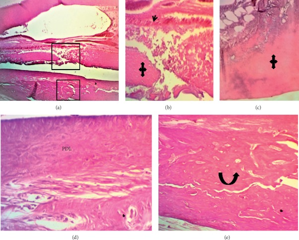
Figure 7.

Microphotographs of longitudinal H&E-stained sections through central incisor after 4 wks of ESRRM putty treatment. (a) The pulpal tissue contained a few long dilated and congested blood vessels, and an irregular deposition of reparative (quad arrows) dentine was seen adjacent to multilayers of short odontoblasts (black arrow head) ((a) 40X; (b-c) 100 X). (d) The periodontal ligament (PDL) had organized dense fibrous principle fibers bundles running parallel to the root surface and inserted in the newly formed bone with minimum congested capillaries and no inflammatory cells infiltration (400X). (e) The gap within the alveolar bone (black curved-up arrow) was filled with compact bone and bone marrow (400X).
