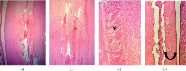
Figure 8.

Microphotographs of an H&E-stained longitudinal section through healthy nonperforated lower central incisors (control group). (a–c) The dental pulp (red arrow heads) showed long dilated congested blood vessels, cellular loose connective tissue, and multiple layers of palisaded columnar odontoblast (black arrow heads) cells adjacent to predentine (40X, 100X, and 200X, respectively). (d) The periodontal ligament (PDL) attached to a thin cementum layer had organized dense fibrous bundles (principle fibers) separated by small areas of vascular connective fibrous tissue. There is no inflammatory cells infiltration. The alveolar bone (black curved up arrow) is compact-dense and contains osteocytes (100X).
