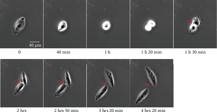
Figure 5.

Cell division and cytodieresis in H28 cells. H28 cells were cultured on a glass surface, and time-lapse was acquired through phase contrast microscopy (dry 20x, DMI 6000 video microscopy, Leica Microsystems). Red arrow indicates the successive steps of cytodieresis residue formation.
