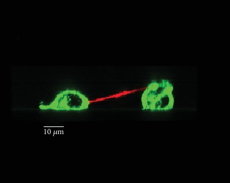
Figure 6.

3D representation of TNT connecting PC12 cells through confocal microscopy. Plasma membrane of living PC12 cells was labeled with Alexa-coupled wheat germ agglutinin (WGA), and Z-stack acquisitions (x, y, z) were performed through confocal microscopy (TCS SP5 X, Leica Microsystems). 3D representation of oblique TNT was obtained through Imaris (Bitplane).
