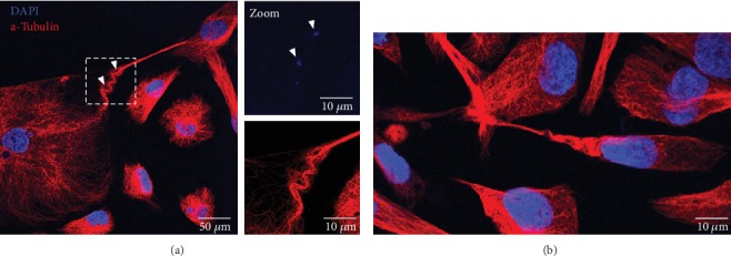
Figure 7.

“Accordion-like shape” of tubulin in TNT1 of HBEC-3 cells (a) and H28 cells (b). HBEC-3 and H28 cells were fixed with PFA 4%; then tubulin (red) was stained through immunodetection (Alexa-546). DNA was labeled with DAPI (blue). Image acquisition was captured with high-throughput confocal microscopy (FluoView FV1000, Olympus™).
