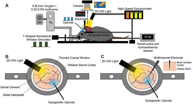
Figure 2.

Chronic imaging preparation. (A) Imaging setup showing inhalational anesthetic maintenance, mechanical whisker stimulation, temperature regulation, hemodynamic imaging (2D-OIS), optogenetic stimulation, and multichannel electrode electrophysiology. (B) First imaging session 2-weeks post-surgery with optogenetic optrode placed over MCA/whisker barrel cortex region. (C) Second imaging session 3-weeks post-surgery with electrode inserted into whisker barrel cortex + optrode for optogenetic stimulation.
