
Figure 7.
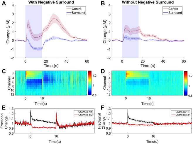
16s photostimulation elicits a negative surround in some SST-ChR2 mice. (A, B) Mean time series showing mean changes in [Hbt] with respect to baseline in SST-ChR2 mice which did (A, n = 9/16 experiments) and did not (B, n = 7/16 experiments) display a negative surround hemodynamic response. Blue box indicates photostimulation. Response in center region shown in red, surround region in blue. (C, D) Mean MUA response in central region across cortical layers for experiments which did (C, n = 9 experiments from 6 mice) and did not (D, n = 7 experiments from 6 mice) display a negative surround hemodynamic response. Color bar represents fractional change. (E, F) Mean time series of response through different cortical layers for experiments which did (E) and did not (F) display a negative surround hemodynamic response. Data shown are mean ± SEM.