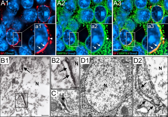
Figure 3.

SP associates with perinuclear endoplasmic reticulum of dentate granule cells. (A1) Higher magnification of a portion of the granule cell layer (gcl). Section immunolabeled for SP (red) and Draq5 (nuclei, blue). Somatic SP is typically found close to the nucleus. (A2) The same section labeled for 78-kDa glucose-regulated protein (GRP78, green), a marker for endoplasmic reticulum (ER), and Draq5 (blue). (A3) Triple labeling of SP, GRP78, and Draq5 demonstrates colocalization of SP and GRP78. Double-labeled structures are abundant close to the nucleus. Note that SP is highly colocalized with GRP78, whereas GRP78 is only colocalized with SP in the perinuclear zone. (a1–a3) Insets show higher magnifications of boxed areas. (B–D) Electron microscopy reveals SP- or GRP78-positive ER cisterns. (B1) Part of a granule cell located in the gcl. Next to the nucleus (N), two SP-positive structures (rectangle; arrow) can be seen. (B2) Higher magnification of rectangle in B1. The DAB precipitate has been intensified with silver particles (see methods). Note association of SP with dense material in between the perinuclear ER. (C) SP was also found in dense material between cytoplasmic ER cisterns, forming triad structures of ER-SP-ER similar to those seen in the spine apparatus or the cisternal organelle. (D1) Granule cell located in the gcl stained for GRP78. Immunopositive ER cisterns were found in the soma and perinuclear. (D2) Boxed area in D1 shown at higher magnification. Arrows point to a cistern connected to the perinuclear ER. Scale bars: 5 μm (A3), 1 μm (inset a3) 500 nm (B1, C), 250 nm (B2, D2), and 1 μm (D1).
