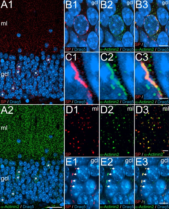
Figure 4.

SP and the Actin-bundling protein α-Actinin2 colocalize in dentate granule cells. (A1, A2) Immunofluorescence labeling for SP (red), α-Actinin2 (green), and Draq5 (blue). Double-labeled SP- and alpha-Actinin2-positive cells are indicated (asterisks). (B) Higher magnification of a somatic SP-positive granule cell in the granule cell layer (gcl) of the dentate gyrus. Somatic SP is primarily found near the nucleus (B1), whereas α-Actinin2 is more widely distributed in the granule cell cytoplasm (B2). The merged picture illustrates a high degree of colocalization between SP and perinuclear α-Actinin2 (B3). (C1-C3) Higher magnification of a portion of the somatic SP-positive granule cell illustrated in (B) demonstrating a perinuclear SP-positive structure, which is also α-Actinin2-positive. (D) Immunofluorescence double-labeling for SP (red, D1) and α-Actinin2 (green, D2) in the molecular layer (ml). The merged image reveals a high degree of colocalization of SP and α-Actinin2 puncta (D3). These puncta represent light microscopic correlates of the spine apparatus organelle. (E) Immunofluorescence labeling for SP (red, E1) and α-Actinin2 (green, E2) in the gcl. The merged image reveals a high degree of colocalization of SP and α-Actinin2 puncta and rods arranged in a string-like fashion (E3). These structures correspond to cisternal organelles in the AIS. Scale bars: 25 μm (A2), 5 μm (B3, E3), 1 μm (C3), and 2 μm (D3).
