Figure 7.

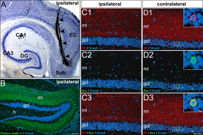
Entorhinal input is necessary for dentate granule cells to express somatic SP or Arc. (A) Horizontal section of a mouse brain showing the wire-knife cut (arrowheads). CA1, CA3, hippocampal subfields CA1, CA3; DG, dentate gyrus; EC, entorhinal cortex; Sub, subiculum. (B) Fluoro-Jade C labeling (green) of degeneration products in the DG (frontal section) ipsilateral to the lesion. Degenerating entorhinal terminals fill the molecular layer (ml) 7 days post lesion. Draq5 (blue) was used as a counterstain to visualize neuronal nuclei. gcl, granule cell layer; h, hilus. (C, D) Portions of the suprapyramidal layer of the DG ipsilateral (C) and contralateral (D) to the lesion site immunolabeled for SP (red) and activity-regulated cytoskeleton-associated protein (Arc, green) and counterstained for Draq5 (blue). Note the absence of SP and Arc-labeling on the side ipsilateral to the lesion (C1-C3; 7 days post lesion) compared to the non-denervated contralateral side (D). The nondenervated contralateral dentate gyrus displays a normal pattern of somatic-SP (D1), Arc (D2), and SP-Arc double-labeled cells (D3). Scale bars: 250 μm (A), 100 μm (B), 80 μm (D3), and 5 μm (D3, inset).
