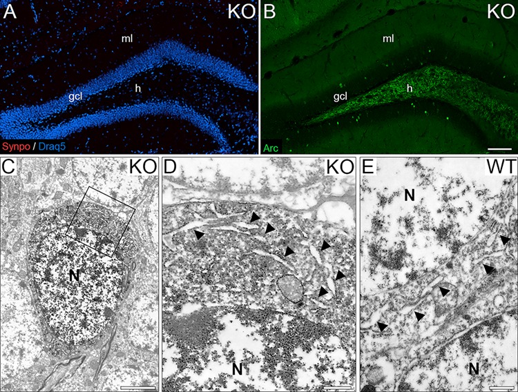
Figure 9.

SP is an essential component of perinuclear ER stacks. (A) Dentate gyrus of a SP-deficient mouse stained for SP. Note the absence of staining for SP in all cellular compartments, that is, somatic SP, dendritic SP, and axonal SP. Draq5 was used as a counterstain to visualize cell nuclei. (B) Dentate gyrus of a SP-deficient mouse stained for Arc. Arc-positive (green) granule cell ensembles were still present in the granule cell layer (gcl) of SP-deficient mice. (C) Electron microscopy of an Arc-positive SP-deficient granule cell. DAB immunoprecipitate is seen in the soma. (D) Higher magnification of boxed area in (C). Cisterns of endoplasmic reticulum (ER) are abundant in the granule cell soma (arrow heads). Neither perinuclear ER stacks nor dense plates were observed (N = 3 animals, 6–7 cells per animal). (E) Somatic SP-negative wild-type (WT) granule cell. ER cisterns (arrow heads) were found throughout the soma but neither perinuclear ER stacks nor dense plates were observed (N = 3 animals, 100 SP-negative cells per animal). gcl, granule cell layer; ml, molecular layer; h, hilus; N, nuclei. Scale bars: 100 μm (A, B), 2.5 μm (C), and 500 nm (D, E).
