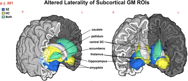
Figure 3.

Subcortical structures with significantly different μLI (P ≤ 0.001) in SZ compared with HC. Color indicates group (SZ—blue, HC—yellow, SZ&HC—green) μLI direction for each ROI. In HC, amygdala, caudate, hippocampus, and nucleus accumbens are left lateralized with thalamus, pallidum and ventral diencephalon being right lateralized. Both a decrease in lateralization (see Table 4; Figure 3) and a reversed trend (i.e., right vs. left lateralization or left vs. right) are noted in SZ for several structures.
