
Figure 1.
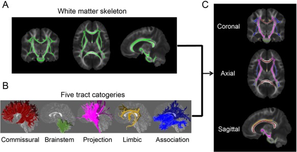
Parcellation of the WM skeleton into five tract groups. (A) The skeleton of the entire WM in coronal (left), axial (middle), and sagittal (right) view after setting the FA threshold 0.2; (B) 3D reconstructed commissural, brainstem, projection, limbic, and association tract groups in sagittal view; (C) parcellation of the WM skeleton into five tract groups with the tract group colors consistent to those shown in (B).