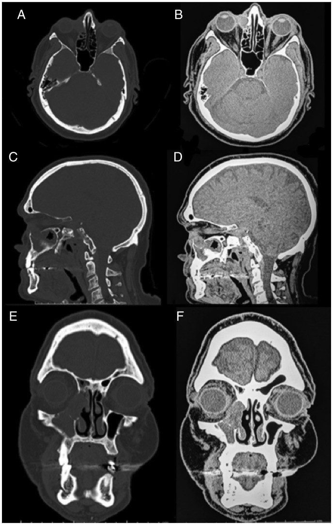
Figure 1.
Noncontrasted maxillofacial computed tomography: (A) axial bone window, (B) axial soft tissue window, (C) sagittal bone window, (D) sagittal soft tissue window, (E) coronal bone-window, and (F) coronal soft tissue window. CT depicts a soft tissue ovoid mass centered in the right nasolacrimal sac, measuring 2.1 × 1.6 × 2.4 cm. No evidence of orbital invasion, but there is extension into the nasal cavity via the nasolacrimal duct to the level of the inferior turbinate and maxillary sinus. Smooth bony remodeling is evident with erosion of the medial lacrimal bone. CT, computed tomography.

