
Figure 3. peBCs Achieve Robust Proximal Epithelial Differentiation in 3 Platforms and More Distal Character in Engineered Lung.
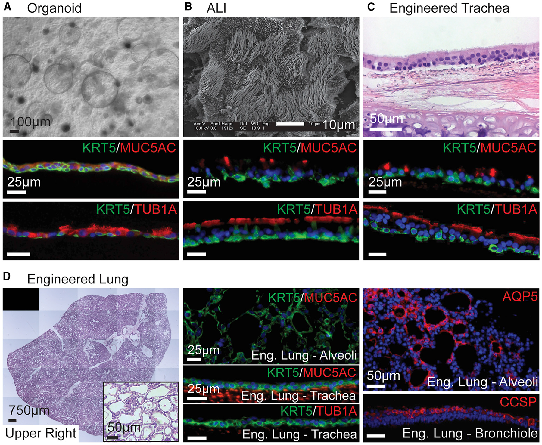
(A–D) peBC differentiated as (A) organoid, (B) ALI, (C) engineered trachea, and (D) engineered lung.
(A) Bright field of organoids at 2 weeks in Matrigel (scale bar, 100 μm).
(B) SEM of ALI at 3 weeks (scale bar, 10 μm).
(C) H&E of engineered trachea at 4 weeks (scale bar, 50 μm).
(D) Stitched H&E image and inset of the upper right lobe of engineered lung at 1 week taken with EVOS system (scale bars 750 μm, 50 μm, resp.); IF of KRT5/ MUC5AC in alveolar region; KRT5/MUC5AC and KRT5/Tub1α in the trachea showing a lack of secretory and ciliated cells (scale bars, 25 μm); IF of AQP5 in alveolar region, CCSP in bronchiole (scale bars, 50 μm).
In (A)–(C), IF of KRT5/MUC5AC and KRT5/Tub1α in organoid, ALI and engineered trachea; organoid lumens and apical cell surfaces oriented up (scale bars, 25 μm).
See also Figures S3–S5.