FIG 2.
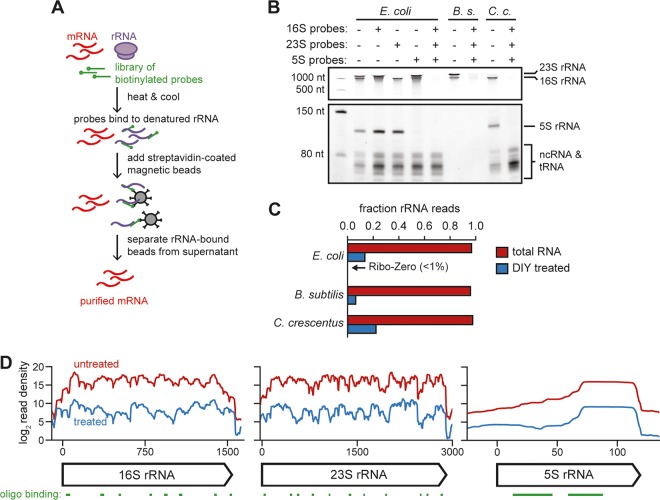
rRNA depletion by oligonucleotide-based hybridization. (A) Cartoon of the rRNA depletion process. (B) Polyacrylamide gel showing total RNA from E. coli, B. subtilis, and C. crescentus before and after rRNA depletion using indicated probe sets. The first lane is a ladder. Approximate positions of abundant RNAs, including rRNAs, are indicated on the right. Note that a lower contrast is shown for the top portion of the gel to resolve 16S and 23S bands. B. subtilis RNA extraction partially depleted the 5S and small ncRNAs (see Materials and Methods). (C) Fraction of total reads aligning to rRNA for rRNA-undepleted and -depleted samples of E. coli, B. subtilis, and C. crescentus total RNA. (D) Summed read counts across the E. coli 16S, 23S, and 5S rRNAs before (red) and after (blue) depletion. The positions of oligonucleotides used for depletion are shown below.

