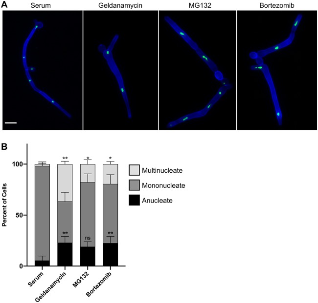
FIG 2.
Filaments formed in response to proteasome inhibition and Hsp90 inhibition share structural similarities. (A) Filaments induced by proteasome inhibition are similar to filaments induced by Hsp90 inhibition but distinct from those induced by serum. Cells were grown under shaking conditions in YPD medium with 10% serum at 37°C for 4 h or in YPD medium with 10 μM geldanamycin, 200 μM MG132, or 520 μM bortezomib at 30°C for 7 h. Cell walls and septa were visualized using calcofluor white (blue), and the nuclei were visualized using a strain with the nucleolar protein, Nop1, tagged with GFP (green). Scale bar, 10 μm. (B) Quantification of the number of nuclei per cellular compartment. The numbers of nuclei in at least 100 cellular compartments were counted for each condition for three biological replicates. Means are graphed with the error bars displaying standard deviations. A paired t test was used to determine significant differences in percentages of multinucleate cells and anucleate cells in response to Hsp90 and proteasome inhibition relative to values for serum-induced filaments. *, P ≤ 0.05; **, P ≤ 0.01; ns, not significant.

