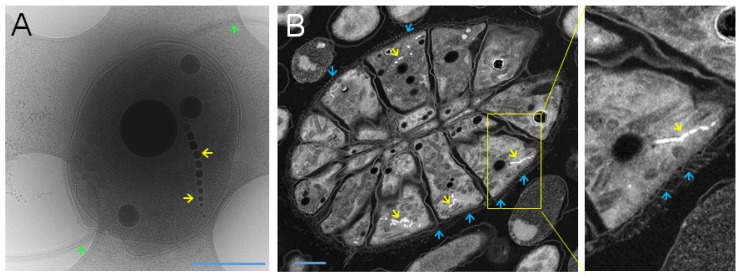
Figure 1.
Magnetosomes and flagella of magnetotactic bacteria. (A) Bilophotrichously flagellated MO-1 cells possess two sheathed flagellar bundles (green arrow) and one magnetosome chain (yellow arrow). (B) Peritrichously flagellated ellipsoidal magnetoglobule with flagella (blue arrows) and magnetosomes (yellow arrows). Only the portion of flagellar filaments in the surface matrix was preserved during sample preparation. Scale bar is equal to 0.5 µm. Courtesy of the electron cryotomography micrograph (A) from Dr. J. Ruan and Professor K. Namba, and of the Scan-TEM high-angle annular dark-field (STEM-HAADF) mode micrograph (B) of ultrathin sections of high-pressure freezing/freeze substitution fixation (HPF/FS) fixed ellipsoidal magnetoglobule from Professor N. Menguy and Dr. A. Kosta.

