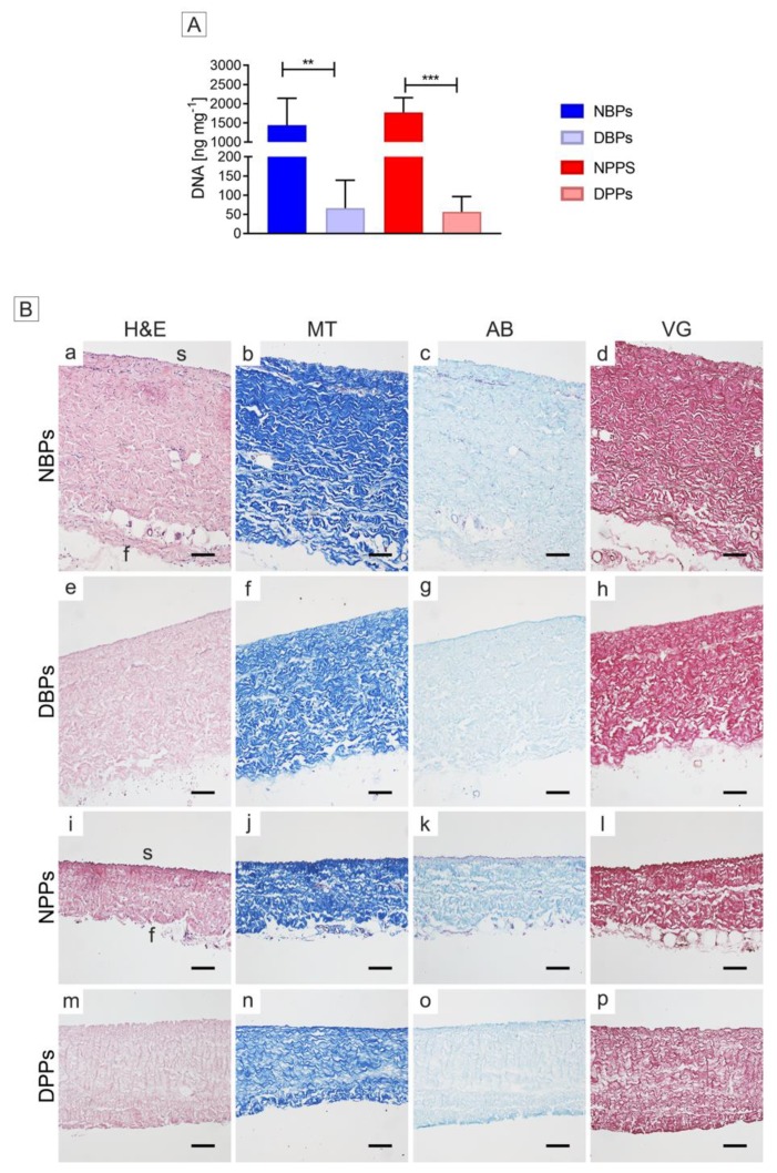
Figure 1.
Decellularization yield of bovine and porcine pericardia after TRICOL procedure. (A) DNA content before and after decellularization. The data are presented as mean ± standard deviation (SD). DNA was significantly reduced in decellularized scaffolds when compared to native ones. **p < 0.01; ***p < 0.001. (B) Histological evaluation before and after decellularization. Intact extracellular matrix (ECM) and no nuclear material were revealed in decellularized bovine (e) and porcine (m) pericardia with respect to their native counterparts (a and i) (Haematoxylin and eosin (H&E)). Collagen organization was intact after decellularization (f for bovine and n for porcine), as in the native corresponding samples (b and j) (Masson trichrome (MT)). A light discoloration was observed for Alcian blue (AB) in decellularized bovine (g) and porcine (o) pericardia with respect to their native counterparts (c and k). No changes in the elastic fibers were detected between the bovine and porcine ECM of native (d and l, respectively) and decellularized (h and p, respectively) pericardia (Van Gieson (VG)). Magnification bars: 100 µm. s= serosa, f= fibrosa. NBP = Native Bovine Pericardium, DBP = Decellularized Bovine Pericardium, NPP = Native Porcine Pericardium, and DPP = Decellularized Porcine Pericardium.

