
Figure 3.
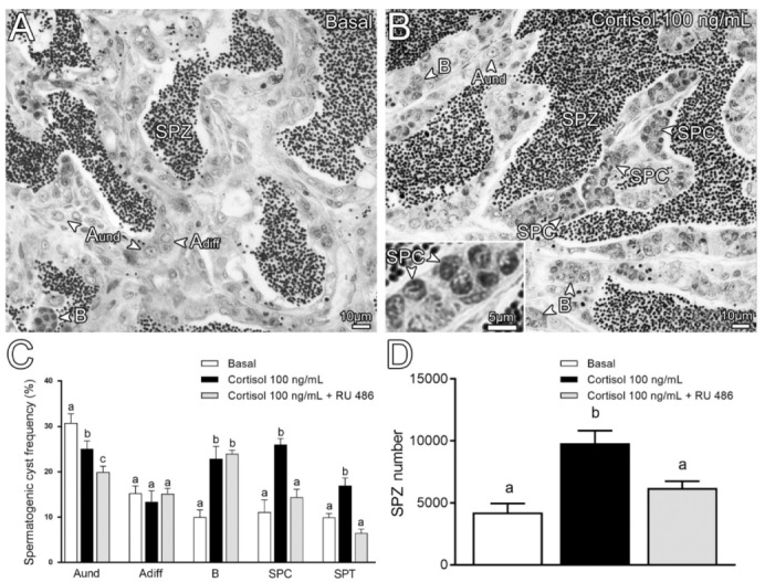
Histological sections from zebrafish testicular explants incubated for 7 days (long-term exposure) with 100 ng/mL cortisol compared with control (basal). (A) Basal (0 ng/mL). (B) Cortisol 100 ng/mL. Several germ cell types are indicated, such as type A undifferentiated spermatogonia (Aund); type A differentiated spermatogonia (Adiff); type B spermatogonia (B), spermatocytes (SPC), and spermatozoa (SPZ). (C) Histomorphometrical analysis of testicular explants incubated for 7 days with cortisol (100 ng/mL) in the absence or presence of 1 µg/mL RU 486 (glucocorticoid antagonist), compared to the control (basal). Bars (mean ± SEM; n = 8) represent the percentage of the spermatogenic cysts at different stages of germ cell development: Aund; Adiff; B; SPC and spermatids (SPT). (D) Spermatozoa quantification per field generated by using IMAGE J Software from zebrafish explants incubated for 7 days with either basal (L-15), cortisol (100 ng/mL), or cortisol (100 ng/mL) + RU 486 (1 µg/mL). ANOVA followed by Student–Newman–Keuls; different letters indicate significant differences (p < 0.05) between different treatment conditions.