
Figure 7.
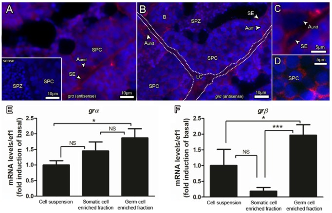
(A–D) Localization of grα (glucocorticoid receptor alpha) expressing cells in the zebrafish testis. Sections were subjected to in situ hybridization using antisense cRNA probe, followed by a fluorescent detection system (HNPP/Fast Red kit (Roche)). The red fluorescence indicates presence of signal, while nuclei were counterstained with DAPI (blue). No specific staining was observed when sections were incubated with sense cRNA probes (inset in (A)). grα mRNA was detected in Sertoli cells (SE), Leydig cells (LC), and several types of germ cells. Type A undifferentiated spermatogonia (Aund); type A differentiated spermatogonia (Adiff); type B spermatogonia (B), spermatocytes (SPC) and spermatozoa (SPZ). The dashed lines delimitate the interstitial compartment. grα (E) and grβ (F) in cell fractions (somatic and germ cell enriched cell fractions obtained from the differential plate method) compared with the total testicular cell suspension. ANOVA followed by Student–Newman–Keuls; *** p < 0.001; * p < 0.05; NS = not significant.