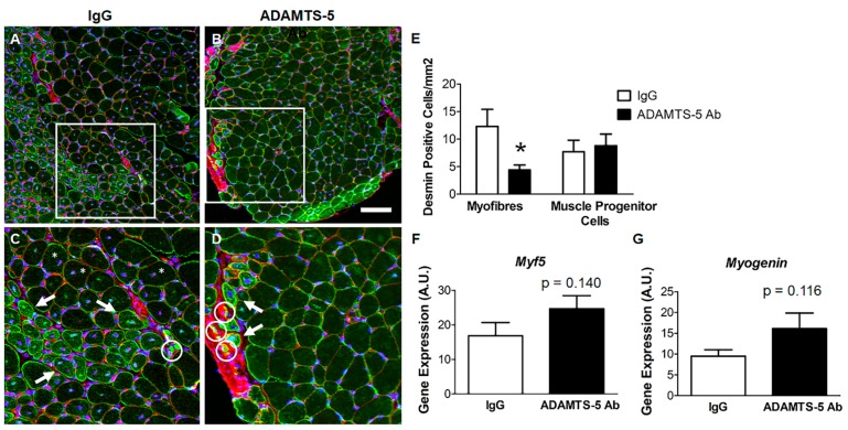
Figure 7.
Markers of regenerative myogenesis in dystrophic EDL muscles following ADAMTS-5 blockade. Desmin immunoreactivity (green) immunoreactivity in EDL cross sections from mdx mice treated with the IgG (A) and the ADAMTS-5 mAb (B); connective tissue (red; WGA) and nuclei (blue). (C–D) Magnified area of interest (white box) with desmin positive muscle progenitor cells (white circles) and desmin positive, newly regenerated myofibers (white arrows); also, not all centrally nucleated fibers had high levels of desmin immunoreactivity (white asterisks). (E) Quantification of desmin positive muscle progenitor cells and regenerating myofibers. (F–G) Effects of ADAMTS-5 blockade on the mRNA transcript abundance of Myf5 and Myogenin. *P < 0.05, independent t-test. N = 8–9 mice for desmin immunoreactivity analysis and n = 13 mice for gene expression analysis. Scale bar = 100 µm.

