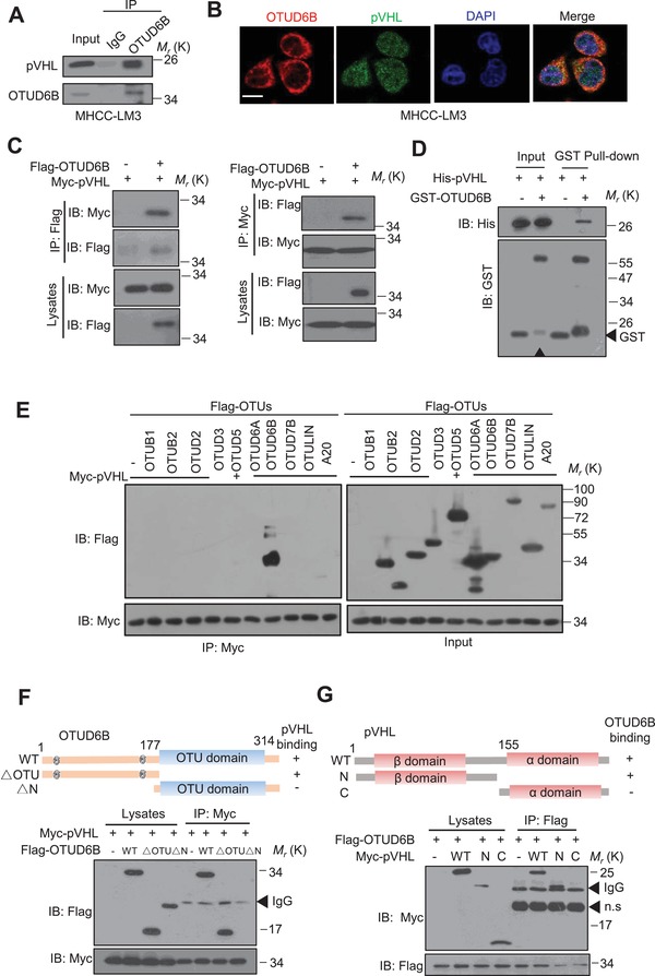
Figure 4.

OTUD6B interacts with pVHL. A) HCC cell lysates were subject to immunoprecipitation with control IgG or anti‐OTUD6B antibodies. B) Immunofluorescence staining was performed in HCC cells with anti‐OTUD6B and anti‐pVHL antibodies to determine the colocalization of these two proteins in MHCC‐LM3 cells (scale bar, 10 µm). C) The lysates of HEK293T transfected with indicated constructs were subject to immunoprecipitation with anti‐myc (or flag) antibody. The immunoprecipitates were then blotted with anti‐flag (or myc) antibody. D) The purified his‐pVHL was incubated respectively with purified GST or GST‐OTUD6B. The mixtures were subjected to GST pull down and blotted. E) The lysates of HEK293T transfected with indicated constructs were subject to immunoprecipitation with anti‐myc antibody. The immunoprecipitates were then blotted with anti‐flag antibody. Overview of E) OTUD6B and F) pVHL structure. HEK293T cells transfected with the indicated constructs were subject to immunoprecipitation with anti‐myc antibody. The lysates and immunoprecipitates were then blotted.
