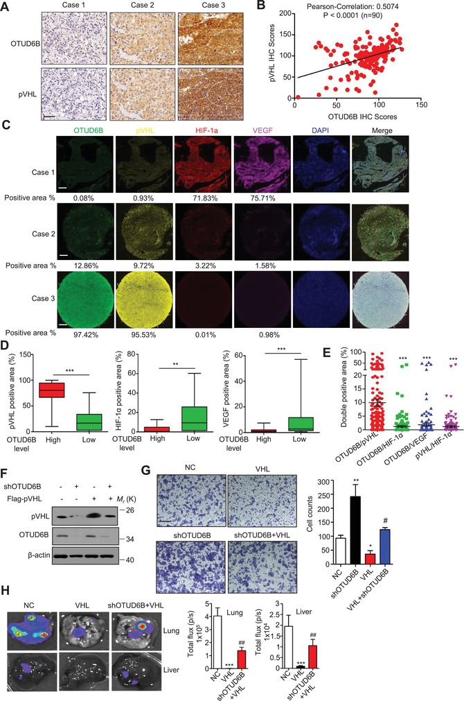
Figure 6.

pVHL mediates the inhibitory effect of OTUD6B on HCC cell migration. A) Representative images from IHC staining of OTUD6B and pVHL in HCC tissues (n = 90) (scale bar, 50 µm). B) The Pearson correlation analysis between OTUD6B level and pVHL level in HCC tissues. C) Representative images from Immunofluorescence staining of OTUD6B, pVHL, HIF‐1α, and VEGF in HCC tissues (n = 90) (scale bar, 300 µm). D) The percentages of pVHL, HIF‐1α, or VEGF positive area in HCC tissues with high or low OTUD6B level were analyzed using the Mann–Whitney U test. * P < 0.05, ** P < 0.01, *** P < 0.001. E) The percentages of double positive area of indicated proteins in HCC tissues were analyzed. * P < 0.05, ** P < 0.01, *** P < 0.001, Student's t test. F) Immunoblotting of pVHL and OTUD6B in HCC cells with stable overexpression or knockdown of indicated genes. G) Transwell assays were performed to identify the capacity of migration in HCC cell with stable overexpression or knockdown of indicated genes. Results are shown as mean ± s.d. n = 3 independent experiments. * P < 0.05, ** P < 0.01, *** P < 0.001, Student's t test. H) 1×10⁶ Luciferase‐expressing MHCC‐LM3 cells were injected into nude mice by tail vein. The mice were euthanized 8 weeks later by a cervical dislocation. Lung and liver tissues were isolated for analysis of IVIS imaging. Results are shown as mean ± s.d. n = 5 independent experiments. * P < 0.05, ** P < 0.01, *** P < 0.001, Student's t test.
